A Solution Trusted by Electron Microscope Manufacturers and Users
What is Electron Microscopy?
Electron Microscopy (EM) is a technique for obtaining high resolution images using a beam of accelerated electrons as a source of illumination. The wavelength of an electron can be up to 100,000 times shorter than that of visible light photons, meaning electron microscopes have a higher resolving power than light microscopes and can reveal the structure of smaller objects.
There are two main types of electron microscope: the transmission EM (TEM) and the scanning EM (SEM). The TEM is used to view thin specimens through which electrons can pass generating a projection image, and the conventional SEM depends on the emission of secondary electrons from the surface of the specimen.
Why Image-Pro?
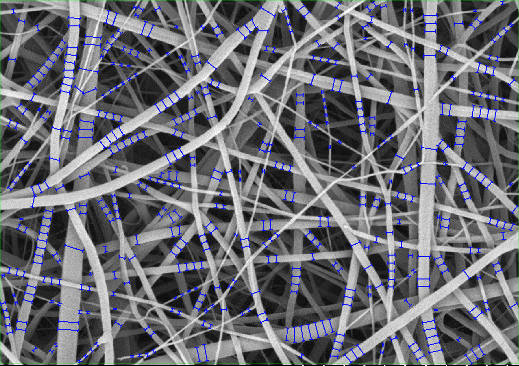
Support 2D images generated by high-res field emission and variable pressure microscopes.
Support 3D images generated by Ion and High kV Transmission microscopes.
Enable an end-to-end solution for all EM customers.
Work with images from nearly all major EM manufacturers.
Provide integrated connection with newer Hitachi systems.
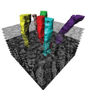
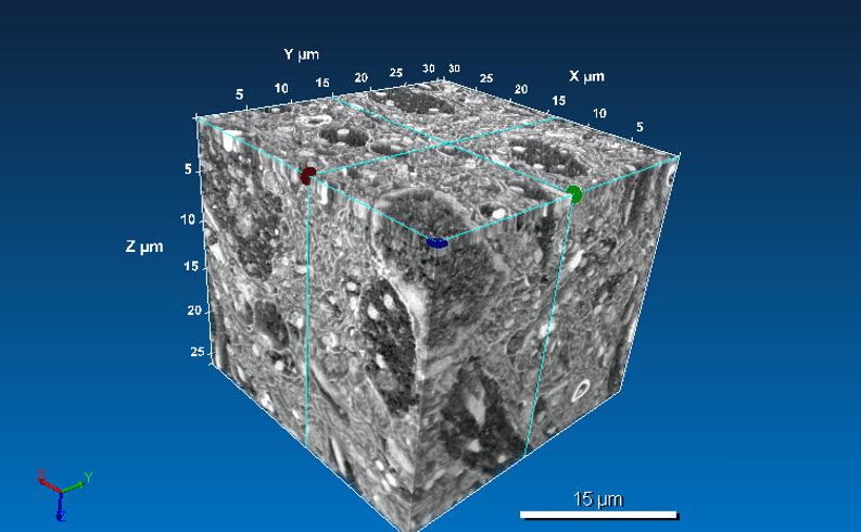
Automatically generate isosurfaces from a collection(s) of 2D semi-automated outlines. Measure volume, surface, intensity, and unique morphological object measurements.
Requires: 3D Visualization Module & 3D Analysis Extension
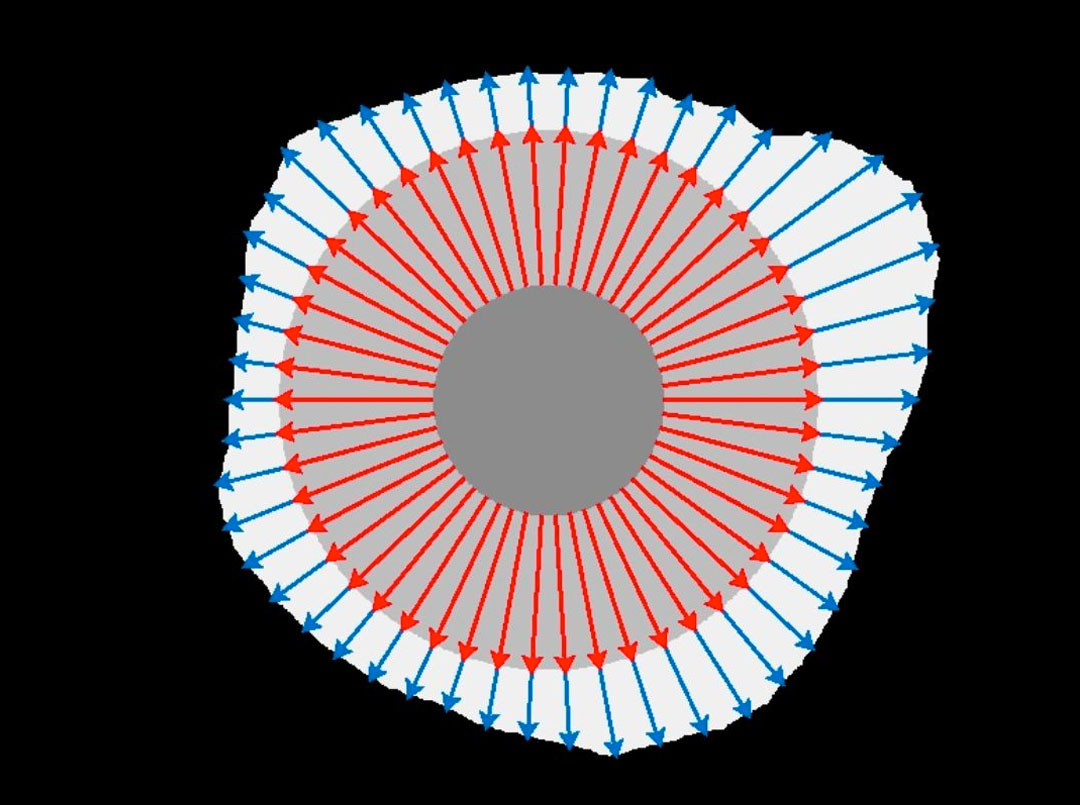
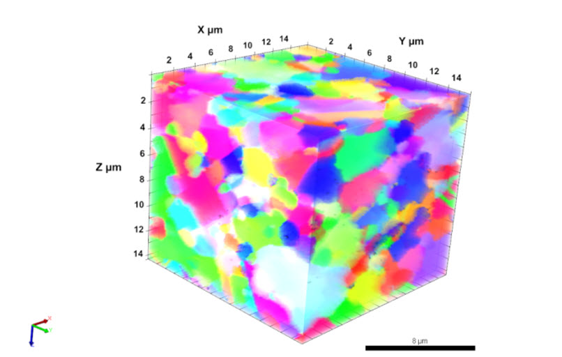
3D-EBSD (Ni) Visualization
Learn More
Visualize EBSD data and the differences in crystalline structure and crystallographic orientation of the grain.
Requires: 3D Visualization Module
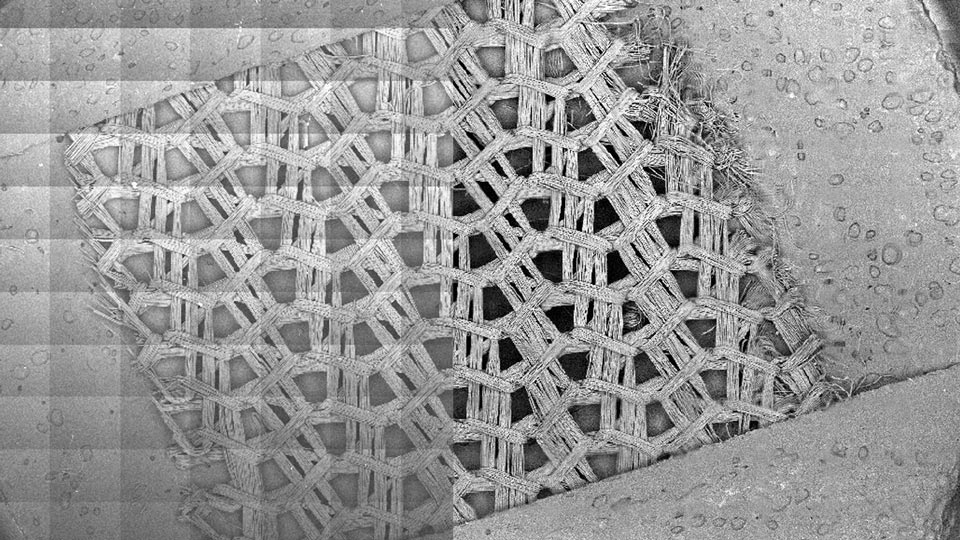
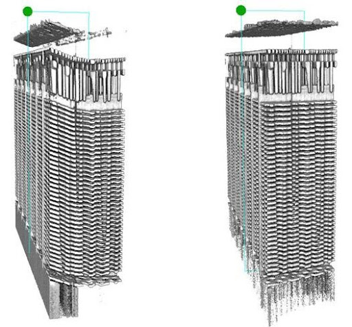
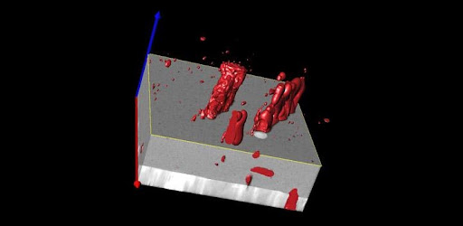
Image Stack Alignment
Learn More
Use automated Fourier or intensity image stack alignment methods to simplify the alignment process of FIB-SEM image stacks.
Requires: 3D Visualization Module & 3D Analysis Extension
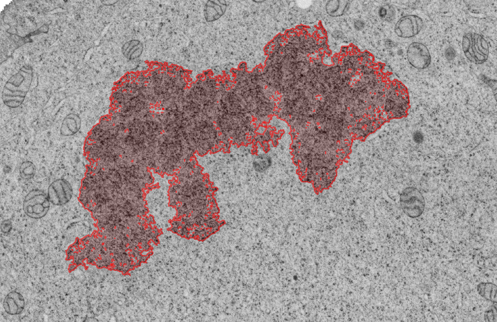
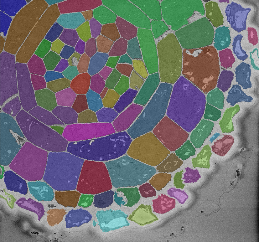
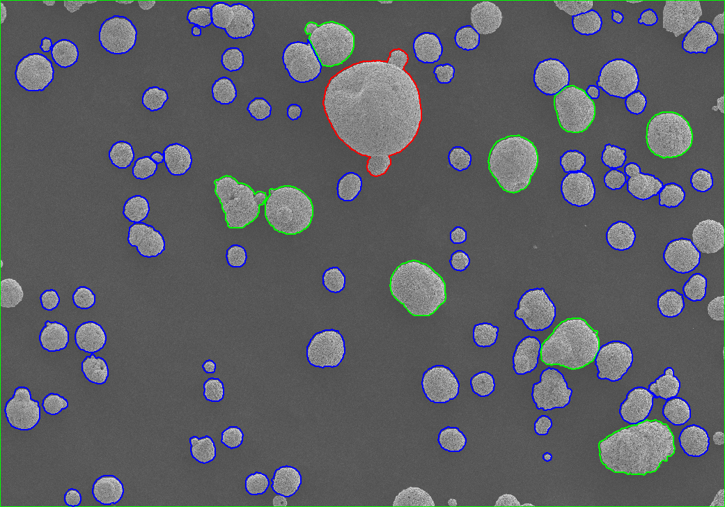
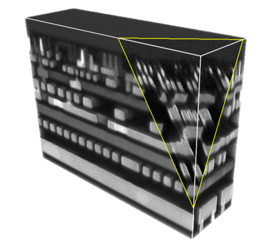
NAND Memory
Learn More
Visualize stacks of FIB-SEM images and unveil internal details using different slicer options.
Requires: 3D Visualization Module
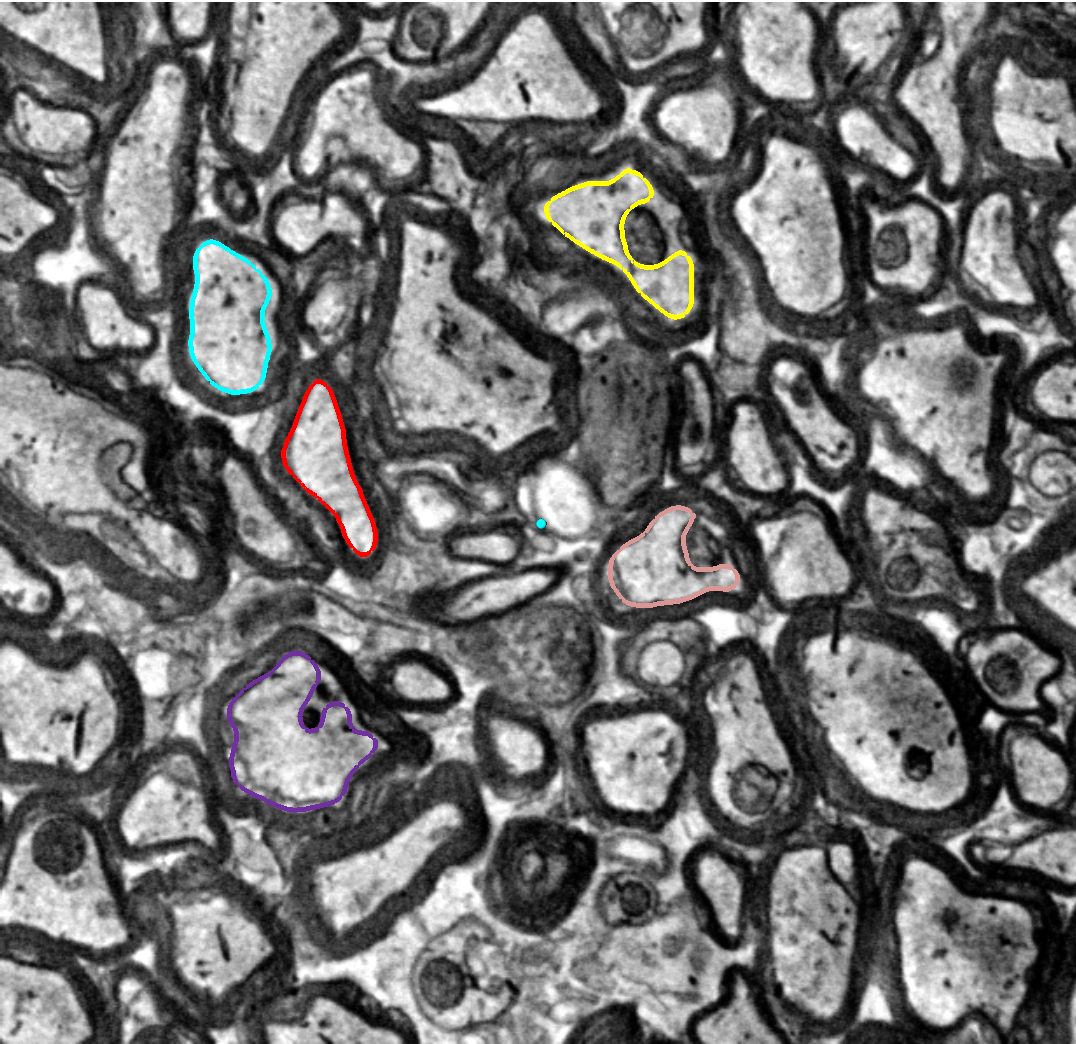
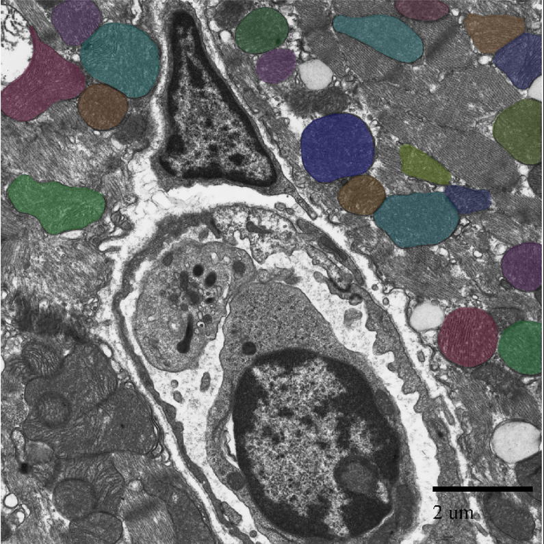
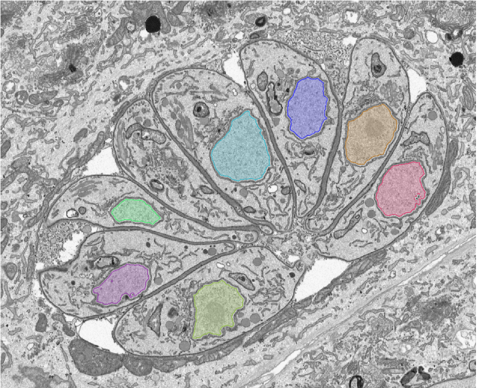
Neuron Analysis
Learn More
Do more than visual Neurons with 3D Segmentation and Guide Segmentation options.
Requires: 3D Visualization Module & 3D Analysis Extension
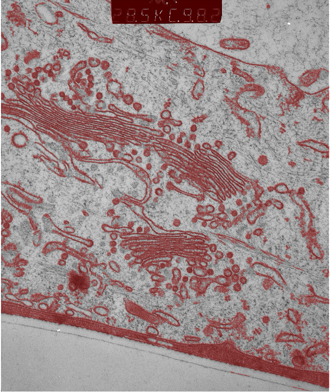
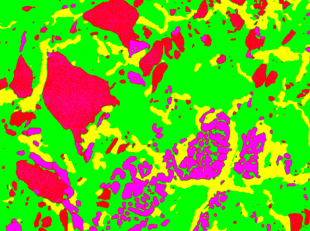
Volume Analysis
Learn More
View, Render, and measure volume data of key elements within your sets. Peel away extra material and spotlight only what is important.
Requires: 3D Visualization Module & 3D Analysis Extension
Image-Pro Has Tools for Your Acquisition Type.
Get Started with Image-Pro for Electron Microscopy