A Solution Trusted by Light Microscope Manufacturers and Users
What is Light Microscopy?
Light Microscopy is a technique for generating magnified images of small objects using visible light and a system of lenses. There are a variety of types including simple and compound lens, stereo, inverted, polarizing, phase contrast, Epifluorescence, Confocal, 2-Photon, and more.
A wide range of illumination techniques are used to generate improved contrast and, in some cases, the addition of standard fluorescent dyes to the sample improve localization of structures and inform about relationships.
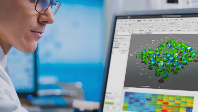
Why Image-Pro?
Supercharge your manual microscopes through powerful capture tools that improve collaboration, handles complex datasets and performs high-volume image analysis. It also optimizes data with filters, coloring, alignment and more all the while saving time on repetitive tasks.
Supercharge manual microscopes by adding powerful capture tools.
Improve collaboration with support for OME.TIF and all major microscope formats.
Handles complex multi-dimensional datasets with ease.
Optimize your light microscope data with filters, coloring, alignment, and more.
Perform expert, high-volume image analysis on nearly any image.
Save time on repetitive tasks using custom macros and a customizable user interface.
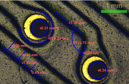
Supported by a wide range of manual measurement and automated measurement features, Image-Pro can quickly be set up to analyze component inspection and measurement.
Perform absolute color measurements or color differences in CIE L*a*b*® or XYZ color coordinates. It’s also just as easy to color correct from a reference image.
Requires: 2D Measurements Module
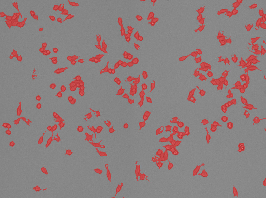
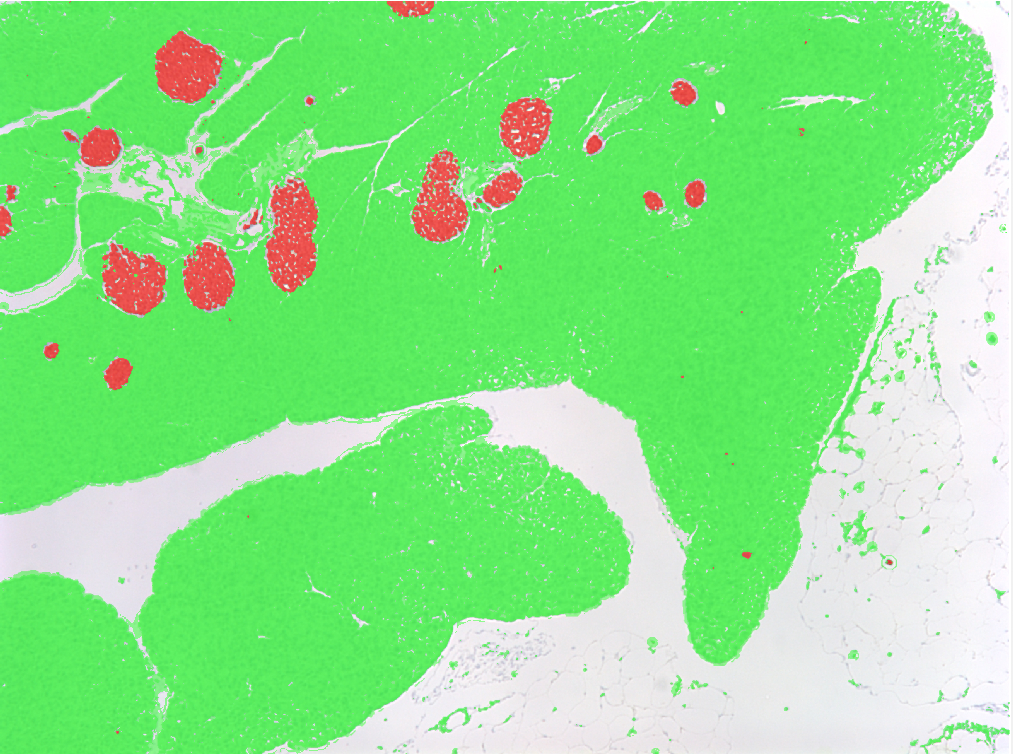
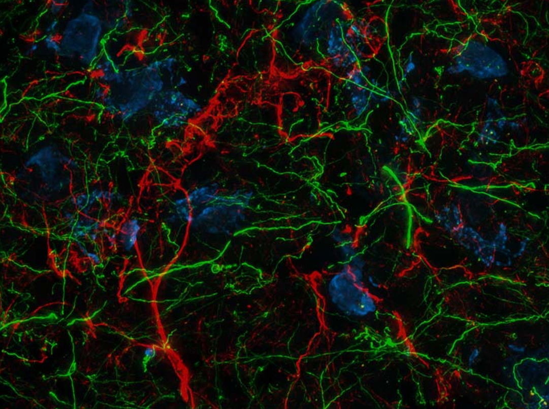
Tissue Stain Analysis
Learn More
Using machine learning-based Smart Segmentation, Image-Pro may be quickly trained to identify stained tissue areas in a reliable and repeatable manner.
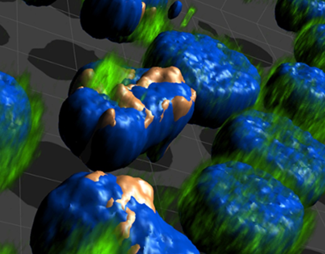
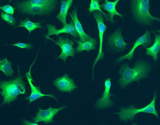
Elevate your capture system to now deliver crisp, high fidelity, fluorescent images during preview and capture.
Required for Live: 2D Capture Module & Real-Time Deconvolution Extension
Required for Post-Acquisition: AutoQuant Deconvolution Module
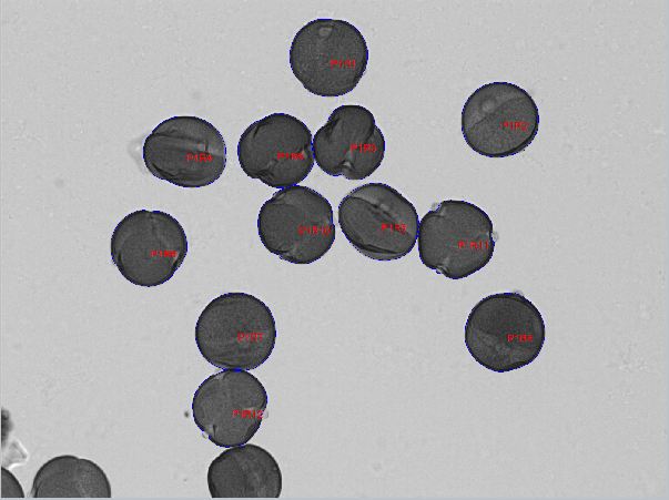
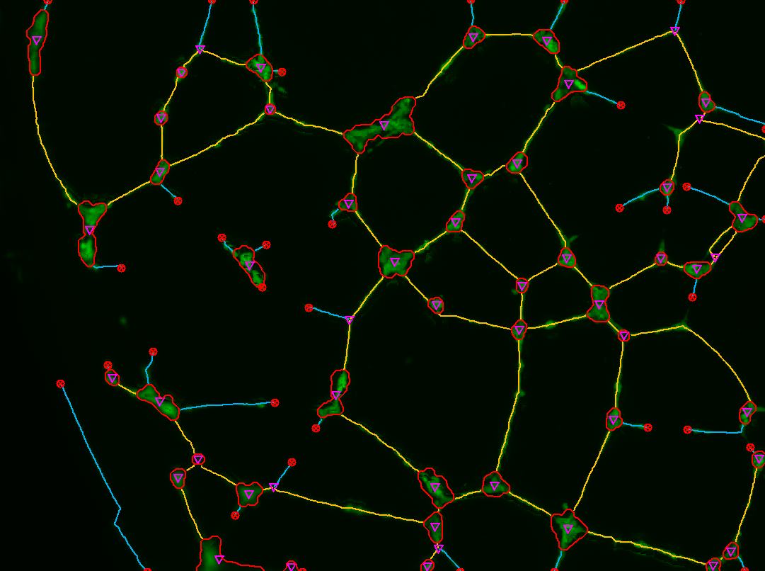
Translocation
Learn More
Measure either the total cellular area of transport of a fluorescently labeled target protein between fluorescently labeled nuclear and cytoplasmic compartments of just the area above threshold.