Cell Morphology (Holotomography)
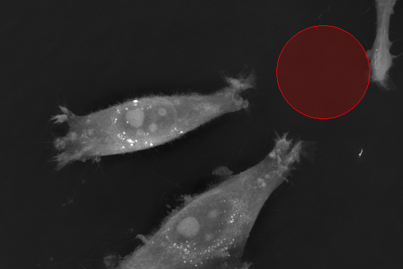
Cellular morphology is highly plastic and dynamic, serving as a critical and informative trait across various biological contexts, particularly in disease research (Tegtmeyer, 2024).
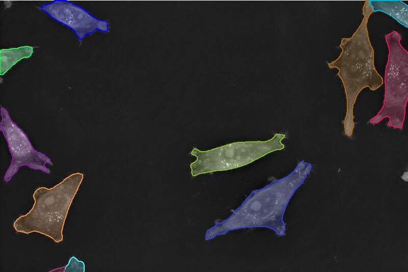
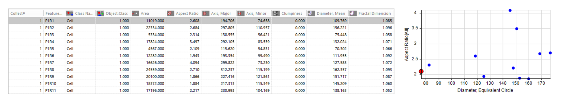
The Image-Pro Cell Morphology (Holotomography) protocol simplifies such studies by providing comprehensive measurements of morphological features at both individual cellular and population levels. This protocol leverages pre-trained deep learning models to ensure accurate assessments of cellular morphology. Additionally, it enables the efficient analysis of large datasets, including complex formats like multi-well plates—even with little to no image analysis experience.
Techniques: Holotomography
How it works


Select Channel
Select the channel that contains cells.
Set Object Diameter
Set the object diameter.
Find Cells
Find cells and nuclei with the Holotomography Cells and Holotomography Nuclei pre-trained deep learning models.
Quantitative results

Automatically generate tables, heat maps, charts and even complex bespoke reports.
Measurement parameters supported
- • Aspect Ratio
- • Axis Major
- • Axis Minor
- • Clumpiness
- • Diameter (Equivalent Circle)
- • Diameter
- • Fractal Dimension
- • Heterogeneity
Solution requirements
Required Modules
Base
2D Automated Analysis
Cell Biology Protocol Collection
Cell Morphology (Holotomography) Protocol
AI Deep Learning
Life Science Models
Fluorescent Cells Model
Recommended Package