Skip links

Mitochondria (TEM)

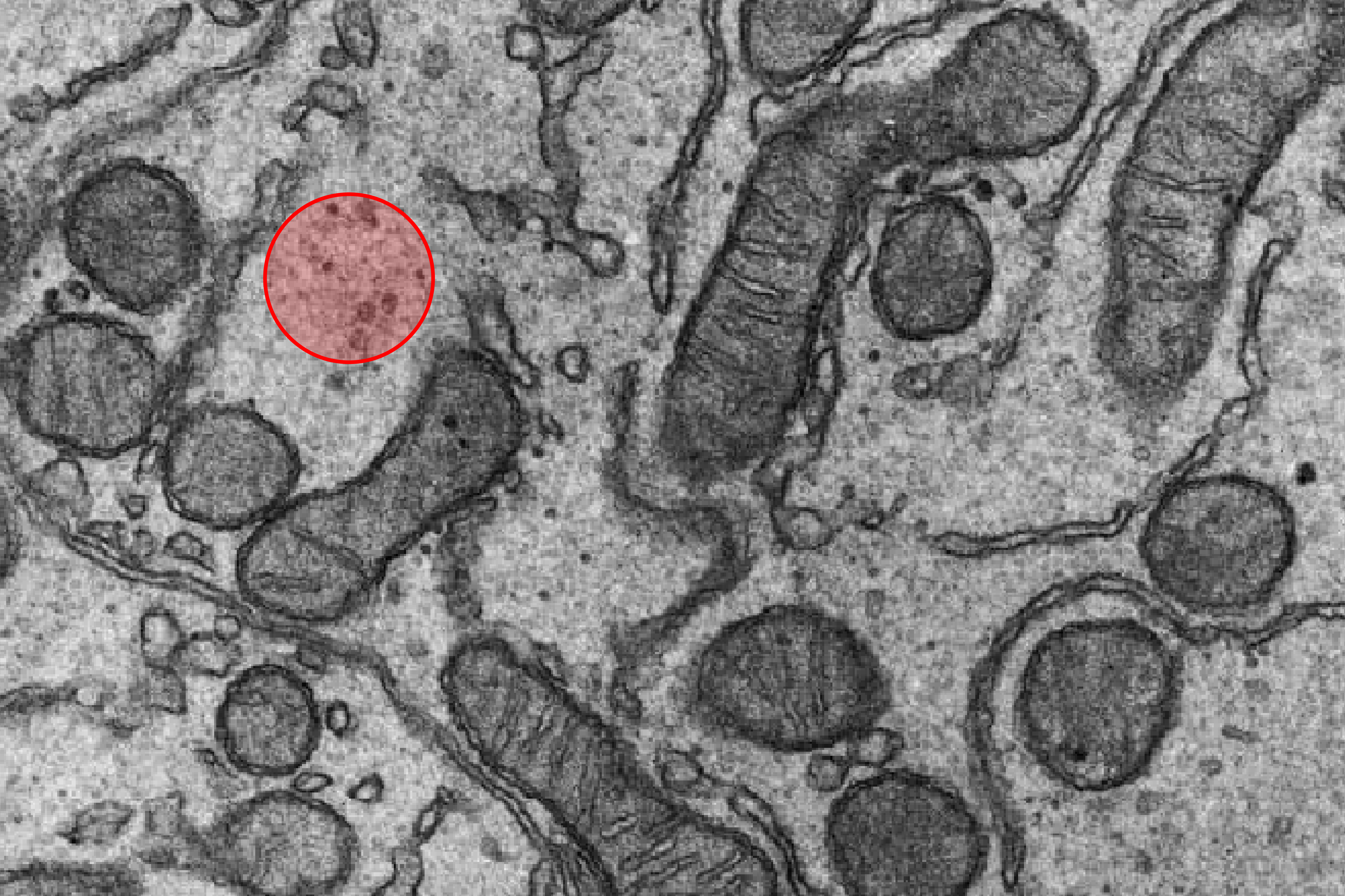
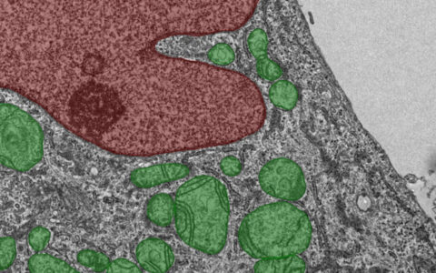
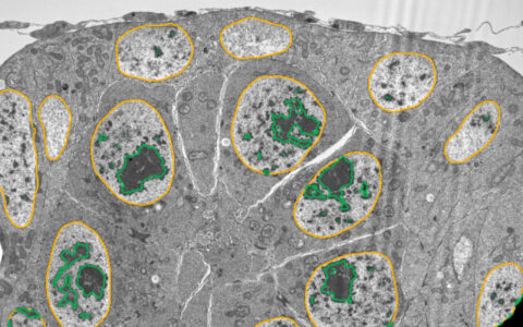
Mitochondria, the powerhouses of eukaryotic cells, are well-recognized in cell biology through textbooks and electron micrographs. However, segmenting mitochondria in electron micrographs has been a significant challenge due to their similar staining characteristics and the variability in their shapes and sizes.

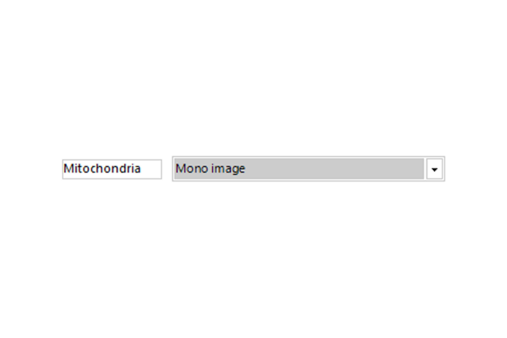
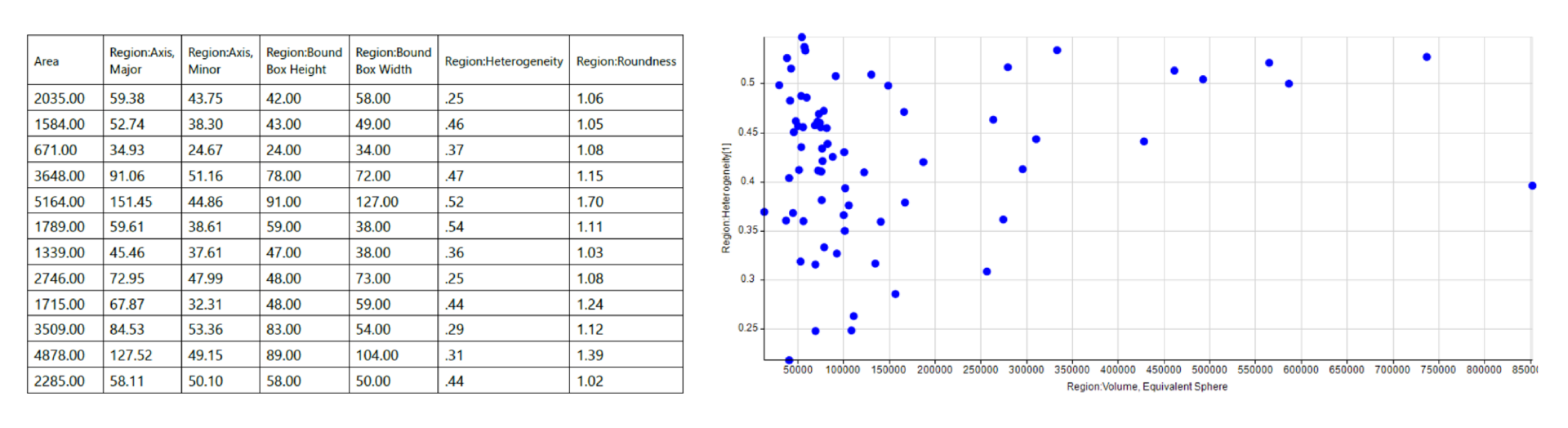
The Image-Pro pre-trained Mitochondria TEM deep learning model addresses this challenge, either used on its own or in combination with the Mitochondria (EM) protocol. These tools simplify the analysis of mitochondria in large volumes of TEM data, allowing for efficient processing with little to no image analysis experience. By leveraging deep learning, the process is made more accessible, ensuring more accurate results for users at all experience levels.

Techniques: FIB-SEM, TEM

How it works

Select Image

Select your TEM image and open the Mitochondria (EM) protocol.

Set Object Diameter

Set the mitochondria diameter.

Find Mitochondria

Find mitochondria with a pre-trained deep learning model.

Quantitative results

Automatically generate tables, heat maps, charts and even complex bespoke reports.

Measurement parameters supported

Solution requirements

Required Modules

Base

2D Automated Analysis

Cell Biology EM Protocol Collection

Mitochondria (EM) Protocol

AI Deep Learning

Life Science Models

TEM Mitochondria Model

RELATED APPS

Request a demo to see how it works

This website uses cookies to improve your web experience.

Download the Mitochondria (TEM) App

Apps are designed for customers with a registered copy of Media Cybernetics software. Please review the App Center specifications tab to ensure your software is supported. To download the App, please fill out the form below.

Download App

Thank you for providing your information.
Please download the App using the link below.

;