Skip links

Nuclei & Chromatin (TEM)

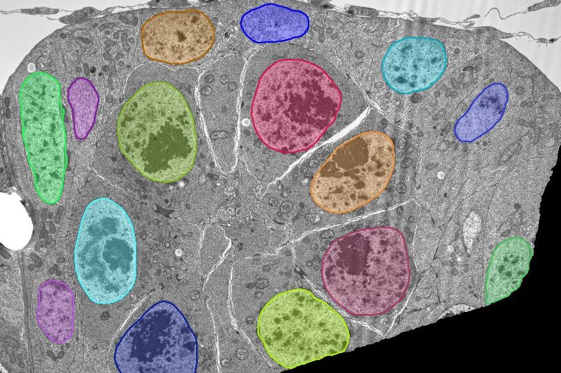
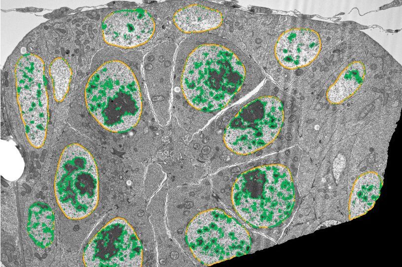
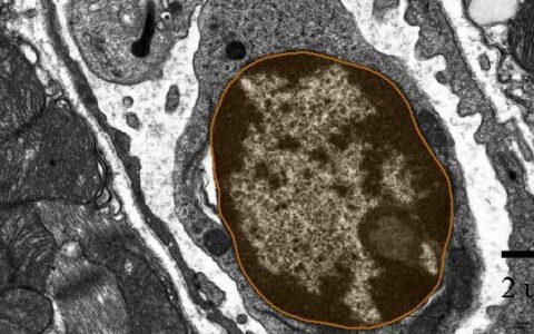
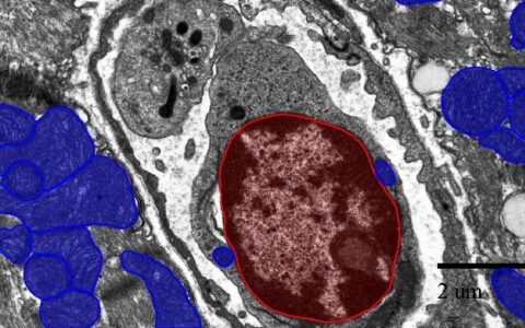
Nuclei are double-membrane-bound organelles that house chromosomes, defining eukaryotic cells. While familiar to all cell biology students through textbooks and electron micrographs, they remain challenging to automatically segment in electron micrographs due to their similar staining characteristics and vast diversity of shapes.

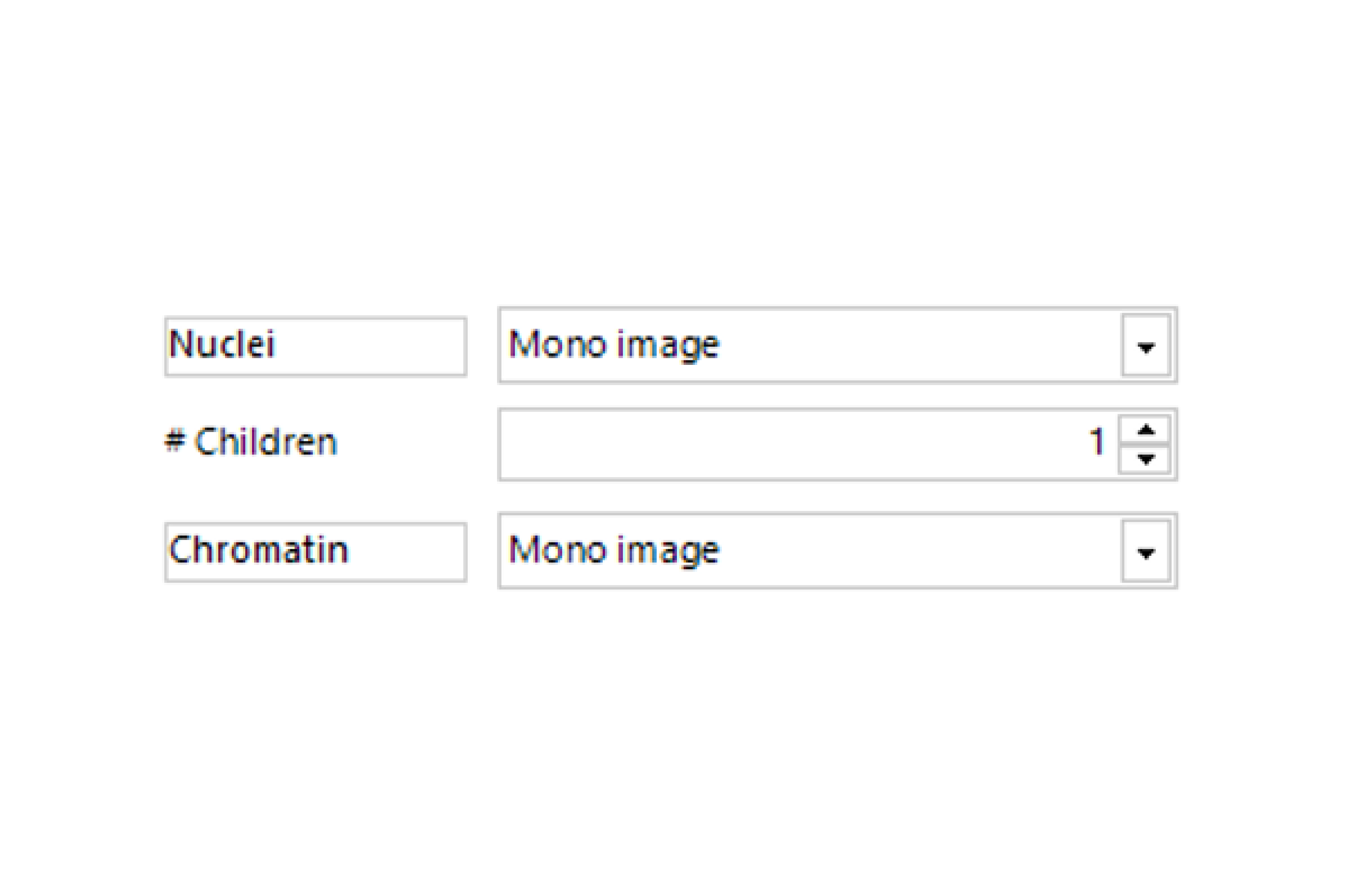
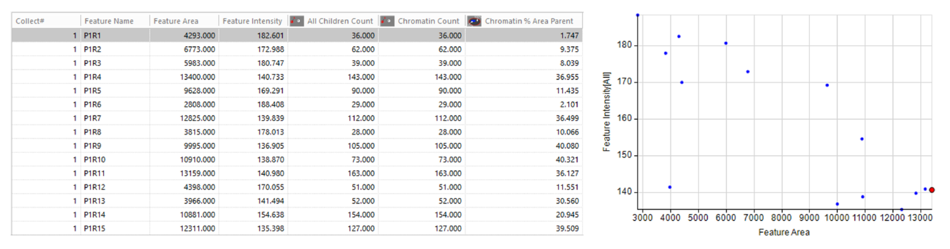
Studies of organelle ultrastructure often rely on the slow and difficult process of manual segmentation (Perez et al., 2014). The Image-Pro pre-trained TEM Nuclei deep learning model overcomes this challenge, working either independently or with the Nuclei and Chromatin (EM) protocol. These tools simplify the analysis of nuclei and chromatin in large volumes of TEM data, requiring little to no image analysis experience.

Techniques: FIB-SEM, TEM

How it works

Select Image

Select your TEM image and open the Nuclei and Chromatin (EM) protocol.

Find Nuclei

Find nuclei with a pre-trained deep learning model.

Find Chromatin

Analyze the contents of nuclei, such as the area occupied by darkly staining chromatin.

Quantitative results

Automatically generate tables, heat maps, charts and even complex bespoke reports.

Measurement parameters supported

Solution requirements

Required Modules

Base

2D Automated Analysis

Cell Biology EM Protocol Collection

Nuclei and Chromatin (EM) Protocol

AI Deep Learning

Life Science Models

TEM Nuclei Model

RELATED APPS

Request a demo to see how it works

This website uses cookies to improve your web experience.

Download the Nuclei & Chromatin (TEM) App

Apps are designed for customers with a registered copy of Media Cybernetics software. Please review the App Center specifications tab to ensure your software is supported. To download the App, please fill out the form below.

Download App

Thank you for providing your information.
Please download the App using the link below.

;