5 Reasons Image-Pro Stands Out for AI-Powered Microscopy Image Analysis
TL;DR — What sets Image-Pro AI Deep Learning apart is how it brings five things together in one tool: multiple proven architectures (Cellpose, StarDist, and BaseUNET) on one on-device Neural Engine, a single microscopist-friendly interface, local-only training and prediction that keeps your images and IP secure, a library of pre-trained models for a fast start, and easy training that needs only a few labeled objects (faster still with an ROI).
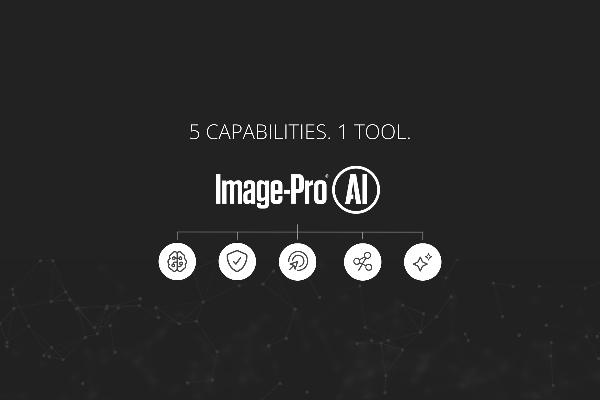
Figure 1. Image-Pro AI Deep Learning combines multiple neural network architectures, local processing, and streamlined training workflows for microscopy image analysis.
Why Microscopy Image Analysis Needs Deep Learning
AI-powered microscopy image analysis has rapidly evolved from an emerging technology into a practical necessity for many laboratories. Traditional image analysis methods based on thresholds, morphology, and edge detection remain valuable, but they often struggle with the complexity of real-world microscopy images.
Touching nuclei, overlapping cells, faint boundaries, noisy images, and variable sample preparation can all require extensive tuning and manual correction. As datasets grow larger, the time required to analyze them can quickly become a bottleneck. Whether you are analyzing fluorescent cells, histology sections, electron microscopy datasets, or materials microstructures, modern AI-powered image analysis software can often solve segmentation problems that would otherwise demand extensive manual intervention.
Deep learning addresses these challenges by learning directly from examples rather than relying on fixed rules. Instead of defining every possible condition, you teach a model what you want to find and let the software recognize similar patterns across new images. Most of these models are built on convolutional neural networks, the architecture behind most modern image segmentation.
If you have read our beginner's guide to AI-powered microscopy image analysis, you already know why deep learning has become one of the most powerful tools available for segmentation. The question today is no longer whether AI works, but what makes one AI image analysis platform better than another. Many software packages can run a neural network. Far fewer make AI practical, secure, and scalable for everyday microscopy workflows. Here are five reasons Image-Pro AI Deep Learning stands apart.
Classical Segmentation vs. Deep Learning Segmentation
Before deep learning became widely available, most microscopy image analysis relied on thresholding, edge detection, watershed segmentation, and morphological filtering. These techniques remain extremely valuable and continue to play an important role in modern workflows.
Deep learning often performs better when images contain overlapping objects, inconsistent staining, weak boundaries, or significant variability. In practice, most modern workflows combine classical image processing and deep learning, using each where it performs best.
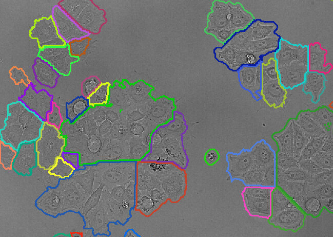
Classical Segmentation
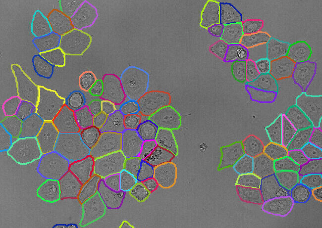
Deep Learning Segmentation
Figure 2. Example of classical segmentation versus deep learning segmentation applied to label-free cell count image analysis.
1. Why One AI Model Isn't Enough for Microscopy
One of the most common misconceptions about AI image analysis is that a single neural network architecture can solve every segmentation problem. In reality, different microscopy applications require different approaches. A model that excels at identifying touching cells may struggle with densely packed nuclei, and a model designed for semantic segmentation may not be ideal for counting individual objects.
This is why experienced image analysts rarely ask which AI model is best. They ask which AI model is best for this particular problem. Many AI tools force users into a single architecture and expect it to perform well across every application. Image-Pro takes a different approach: at the core of the software is the Image-Pro Neural Engine, which supports multiple deep learning architectures through one consistent workflow and interface.
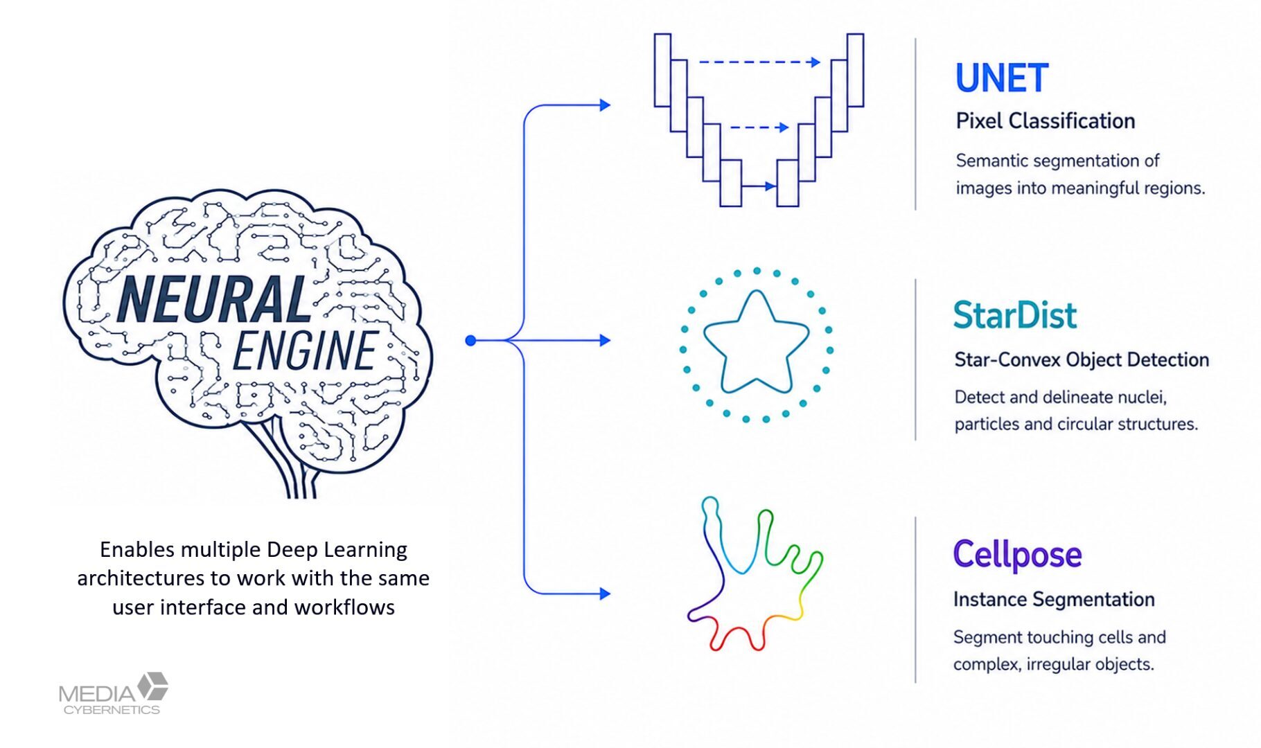
Figure 3. The Image-Pro Neural Engine supports multiple segmentation architectures through one consistent workflow.
Cellpose
Instance segmentation for cells, particles, fibers, and touching objects. See Stringer et al., Nature Methods, 2020.
Figure 4. Before-and-after example of Cellpose deep learning segmentation for fiber thickness analysis in electrospun EM imaging.
StarDist
Instance segmentation optimized for nuclei, grains, and rounded structures. See Schmidt et al., 2018.
Figure 5. Before-and-after example of StarDist deep learning segmentation for overlapping nuclei in cell biology imaging.
BaseUNET
Semantic segmentation for tissue regions, material phases, and area measurements, based on the U-Net architecture (Ronneberger et al., 2015).
Figure 6. Before-and-after example of BaseUNET deep learning segmentation for particle phase imaging.
Rather than forcing you to adapt your science to a single model, Image-Pro lets you choose the architecture that best matches your biological or materials science question. If you want a refresher on the distinction between these model types, see our primer on semantic vs. instance segmentation.
Cellpose vs. StarDist vs. BaseUNET:
Which Model Should You Choose?
One advantage of the Image-Pro Neural Engine is the ability to select the architecture that best matches the scientific problem. For many users, Cellpose provides an excellent starting point because of its versatility. StarDist often performs exceptionally well on crowded nuclei and similarly shaped structures, while BaseUNET is typically preferred when measuring area coverage rather than identifying individual objects.
The important takeaway is that no single architecture is ideal for every microscopy problem, because different scientific questions require different approaches. For more on choosing between object-level and pixel-level methods, see our guide to semantic vs. instance segmentation.
2. Why Data Security Matters in AI-Powered Microscopy
As AI becomes more common in scientific workflows, many solutions have moved training and prediction into the cloud. For some applications that is perfectly acceptable, but for others it introduces significant concerns.
Researchers may be working with sensitive data, including:
- Proprietary materials
- Unpublished research
- Confidential client projects
- Regulated workflows
- Sensitive intellectual property
Uploading images to external servers can create uncertainty around data ownership, security, and compliance. Image-Pro was designed with a different philosophy: all training and prediction occur locally on your workstation.
Because the Image-Pro Neural Engine operates on-device:
- Images remain inside your organization
- No cloud uploads are required
- No third-party servers store your data
- Performance is not dependent on internet speed
This approach is particularly valuable when working with proprietary research, regulated workflows, or confidential client data, and it eliminates the delays associated with uploading large microscopy datasets. For many laboratories, keeping data local is not simply a preference. It is a requirement.
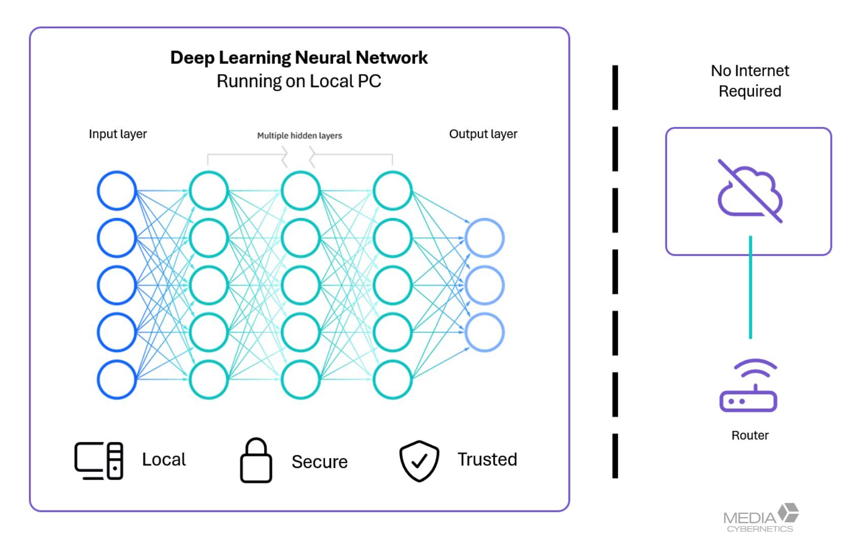
Figure 7. Training and prediction occur locally, helping keep microscopy images and intellectual property under your control.
3. Why AI Adoption Often Fails in the Laboratory
When scientists evaluate AI software, accuracy is rarely the only challenge. Usability often becomes the larger obstacle. Many deep learning platforms require users to understand training parameters, model architectures, configuration files, and coding environments before they can generate useful results. Most microscopists do not want to become machine learning engineers; they simply want accurate answers from their images, and Image-Pro was designed around that reality.
The same workflow handles prediction, fine-tuning, and training through a single, consistent interface, so you move from a first result to a refined model without ever leaving the tool or writing a line of code.
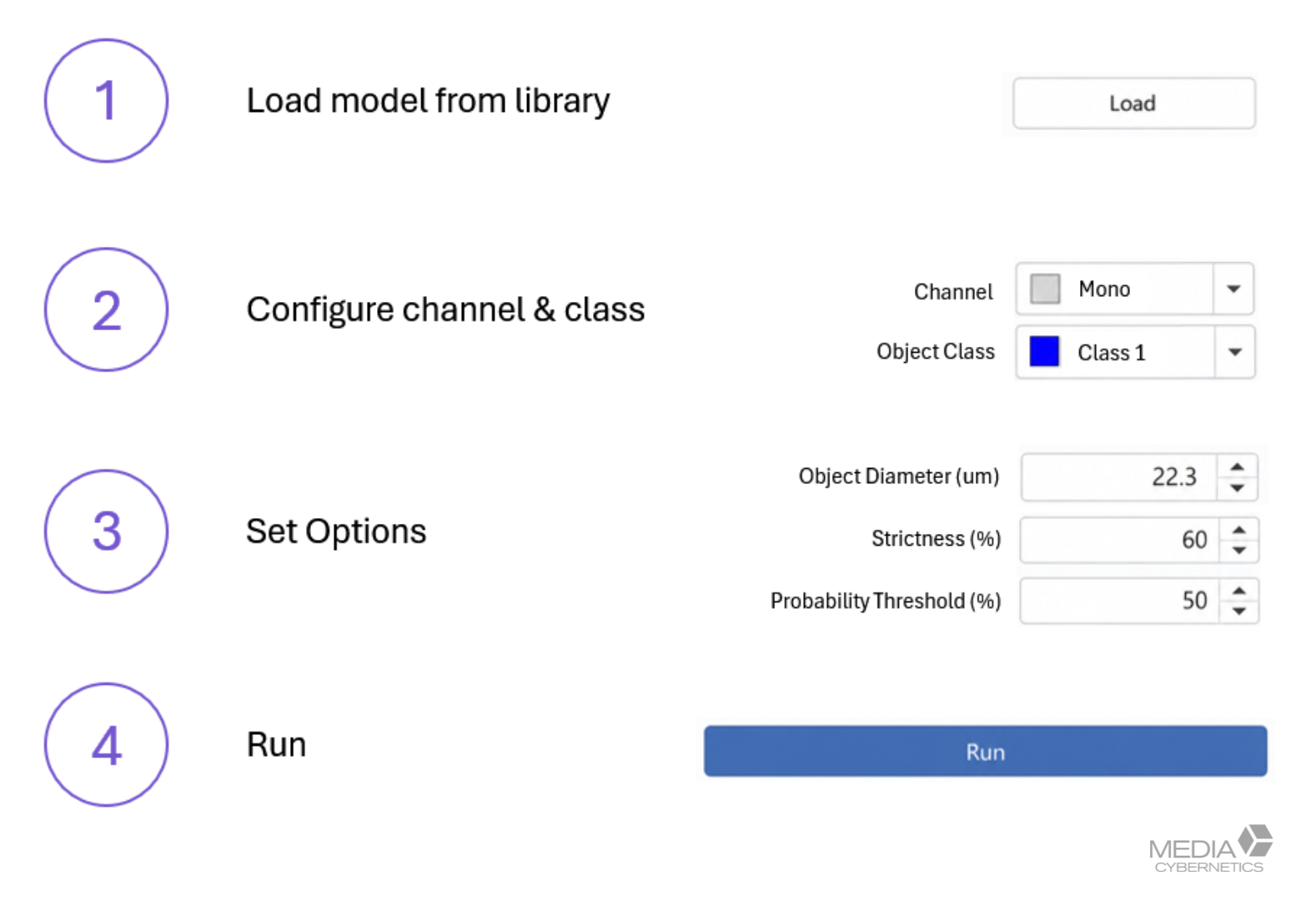
Figure 8. Prediction, fine-tuning, and training are managed through a workflow designed specifically for microscopy users.
Real-World Impact
The AI Deep Learning tool is flexible and efficient, with pre-trained models that provide a strong starting point for new projects. It has significantly improved our productivity and reduced the time required to generate reliable segmentation results.
Dale K. Purcell, PhD
President at Chemical Microscopy LLC
Prediction itself requires only a handful of intuitive controls, such as object size estimation, confidence thresholds, and scale adjustment. You can quickly generate results, review performance, make corrections, and retrain models without writing code. The result is a platform that stays approachable for new users while still giving experienced researchers the flexibility they need.
4. Why Starting from Scratch Is Usually Unnecessary
One of the biggest misconceptions about deep learning is that every model must be trained from the beginning. In practice, many successful AI workflows start with a model that already understands a specific type of object, which dramatically reduces the time required to achieve useful results.
Image-Pro includes a growing library of pre-trained models for both life science and materials science applications. You can explore many of these ready-to-use solutions in the Image-Pro App Center.
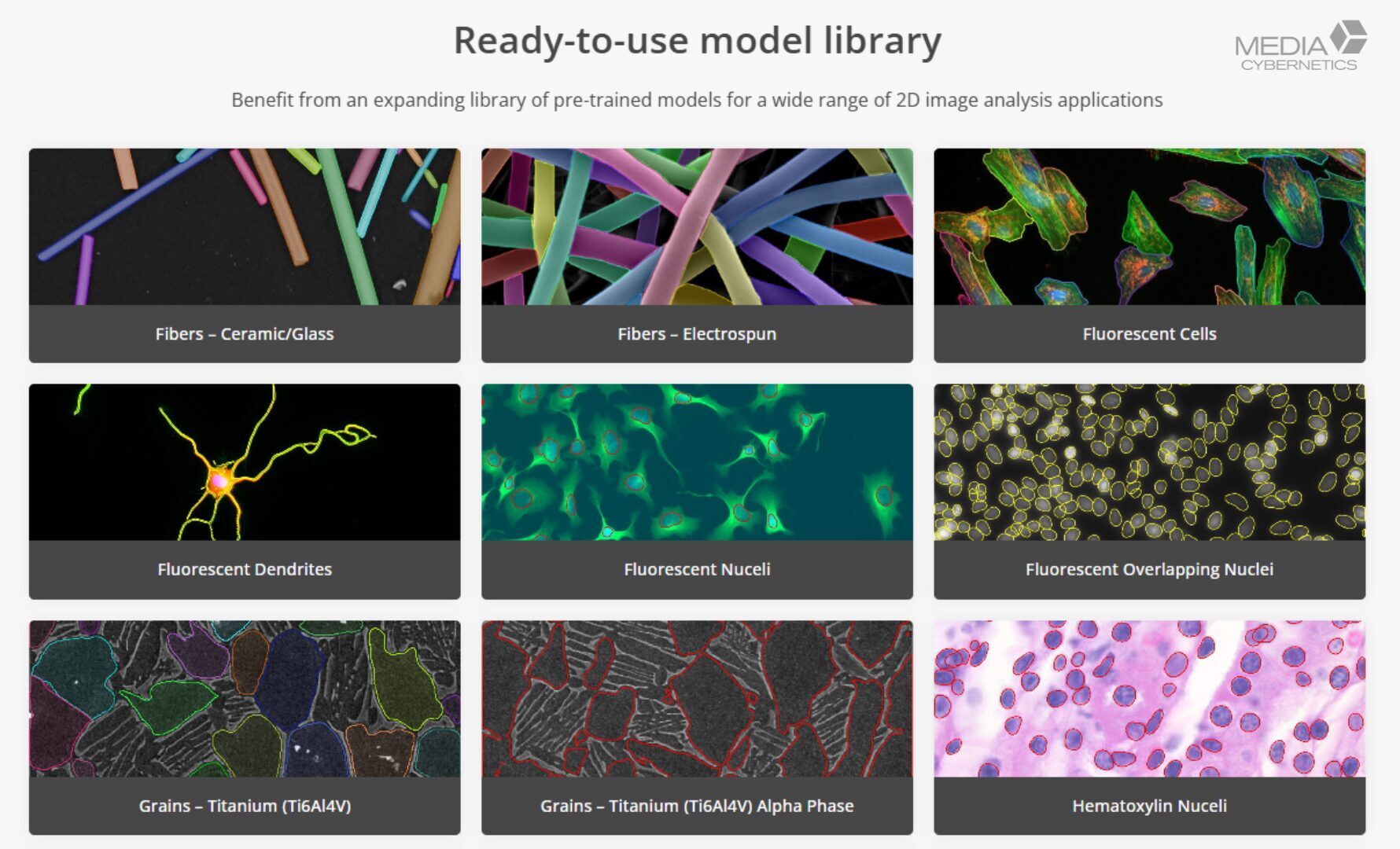
Figure 9. Built-in pre-trained models help users reach accurate results faster.
Life Science Examples
- Fluorescent nuclei
- Overlapping nuclei
- Label-free cells
- Holotomography cells
- Retinal blood vessels
- TEM mitochondria
- TEM nuclei
Materials Examples
- Ceramic fibers
- Glass fibers
- Electrospun fibers
- Titanium grains
- Alpha-phase grains
- Particles
- Widmanstätten structures
Many users can achieve meaningful segmentation immediately without any additional training. Even when a model is not a perfect fit, it often provides an excellent starting point for further refinement.
5. Why Training an AI Model Can Be Easier Than You Think
Many researchers assume successful AI training requires thousands of annotated objects and weeks of preparation. In reality, modern fine-tuning workflows can be surprisingly efficient.
Image-Pro follows a straightforward, human-in-the-loop process:
1. Predict
2. Correct
3. Train
4. Repeat
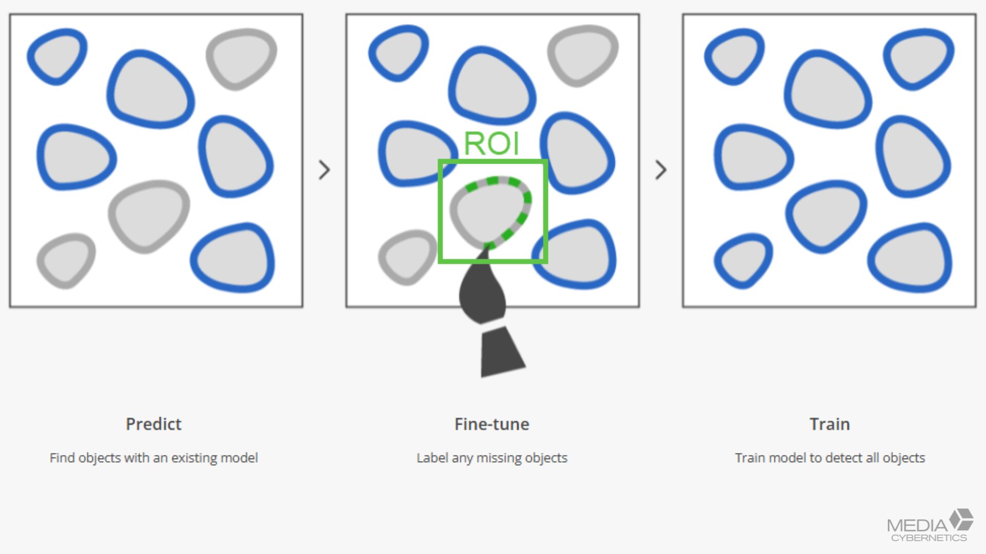
Figure 10. A practical workflow for improving segmentation accuracy with minimal labeling effort.
Starting with a pre-trained model dramatically reduces the amount of annotation required, and you can accelerate training further by labeling only carefully selected regions of interest (ROIs) rather than entire images. One rule of thumb matters here: within whatever region you label, be sure to label every object, since anything left unmarked is treated as background. Once a model performs well, you can incorporate it directly into Analysis Protocols for automated, repeatable workflows. This approach lets you improve performance while minimizing the time spent creating training data.
Bringing It All into Focus
"Remarkably Accurate. Delightfully Efficient." is the promise behind Image-Pro's AI Deep Learning, and these five differentiators are how it delivers on both halves of that line. The accuracy comes from running multiple proven architectures through one engine. The efficiency comes from a microscopist-friendly interface, a head start from pre-trained models, and training that asks for only a small number of labeled objects. The on-device design ties it together by keeping your data and your IP secure and your results fast.
The result is deep learning you can actually put to work: matched to your question, easy to operate, safe with your data, and quick to get to a dependable answer.
Final Thoughts
Choose the right AI for the job with multiple architectures, including Cellpose, StarDist, and BaseUNET.
Keep your data secure with local training and prediction that never requires cloud uploads.
Get started faster using built-in pre-trained models for life science and materials applications.
Train with less effort through efficient fine-tuning workflows and ROI-based labeling.
Scale your analysis by integrating trained models directly into Image-Pro Analysis Protocols.
Frequently Asked Questions
The best solution depends on your application, but you should look for software that combines accurate segmentation models, workflow automation, ease of use, and secure data handling. Image-Pro AI Deep Learning combines multiple deep learning architectures, local processing, pre-trained models, and analysis protocols within a single platform.
Cellpose is often an excellent starting point for cell segmentation because it performs well across many biological image types. StarDist frequently performs better on crowded nuclei, while BaseUNET is preferred for semantic segmentation tasks involving area measurements and coverage analysis.
Cloud-based AI may be appropriate in some environments, but many organizations have concerns related to intellectual property, unpublished research, regulatory compliance, and data security. Image-Pro performs prediction and training locally on your workstation, helping keep microscopy data under your control.
Yes. Deep learning can learn complex visual patterns that are difficult to capture with traditional threshold-based approaches, and it often performs significantly better on noisy, low-contrast, or crowded microscopy images.
Many workflows can achieve meaningful improvements using a relatively small number of carefully labeled examples, particularly when starting from a relevant pre-trained model.
Yes. Models can be incorporated directly into Image-Pro Analysis Protocols, enabling repeatable and scalable image analysis across experiments, users, and datasets.

Media Cybernetics
sales@mediacy.com
Related Links