- Products
- Features
- App Center
- Resources
Self-Service Support
Live Support & Services
- Company
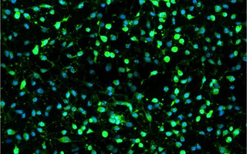
Cell viability assays measure the proportion of live and healthy cells in a culture, often used to screen cell populations' response to drugs or chemical agents (Kamiloglu et al., 2020). Cell viability can be assessed by staining live cells with dyes such as Calcein-AM (which is converted to a fluorescent form in living cells) or by detecting dead cells with stains like Propidium Iodide (which is excluded from healthy cells but stains the DNA of dying or dead cells). These assays can even measure both live and dead cells simultaneously.
The Image-Pro Live/Dead Cell Viability protocol is compatible with different combinations, including a) live and total cells, b) dead and total cells, or c) live, dead, and total cells. Pre-configured versions of the protocol are available for these combinations, making it easy to analyze large datasets in complex formats, such as multi-well plates, with minimal image analysis experience.
Techniques: Fluorescence
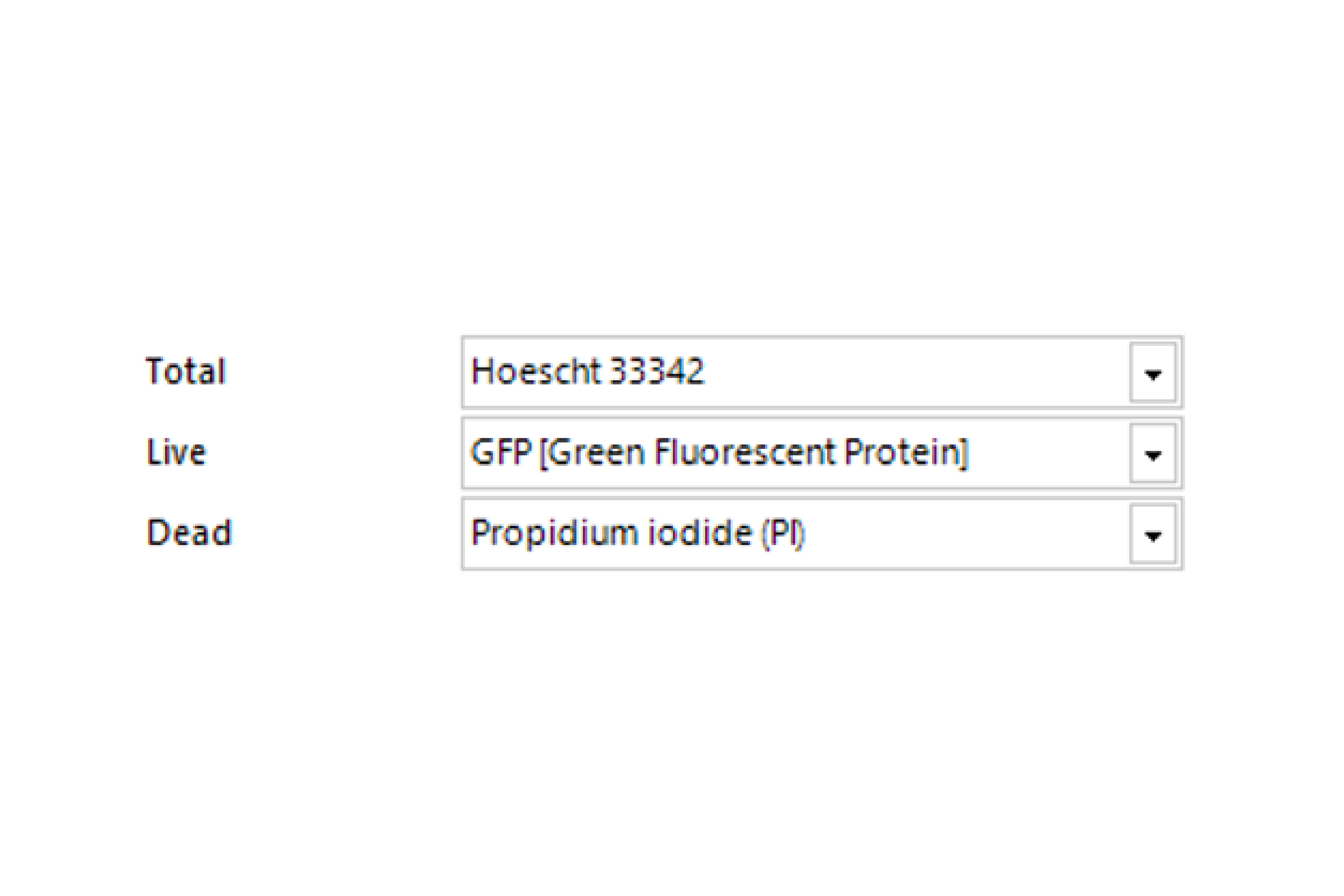
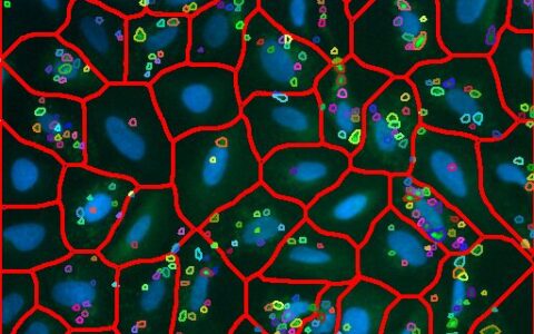
Select Channel
Select the channels that contain total cells, labeled live cells, and labeled dead cells.
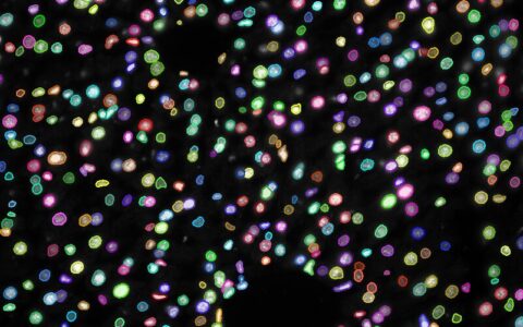
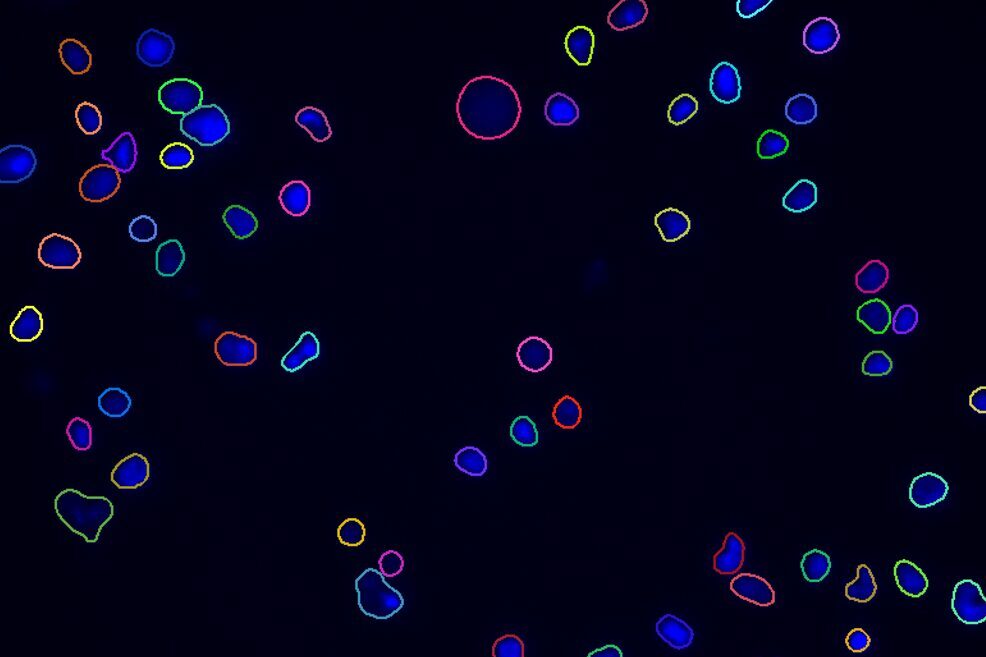
Find Cells or Nuclei
Find cells or nuclei with a pre-trained deep learning model, machine learning, or threshold segmentation.
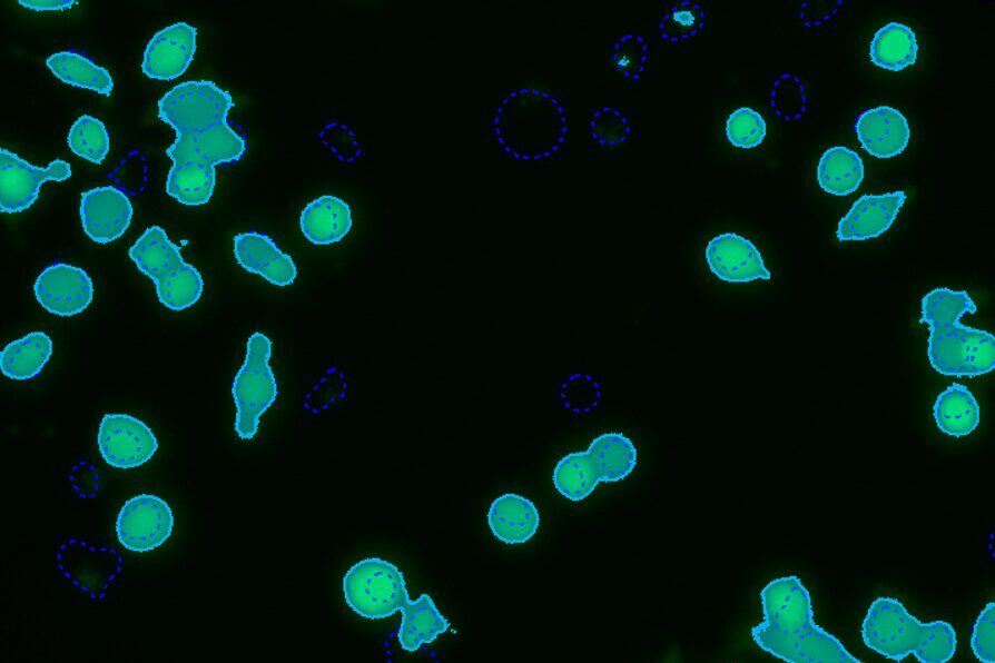
Find Live/Dead Cells
Identify cells that are labeled as ‘live’ or 'dead' with either threshold segmentation or machine learning segmentation.
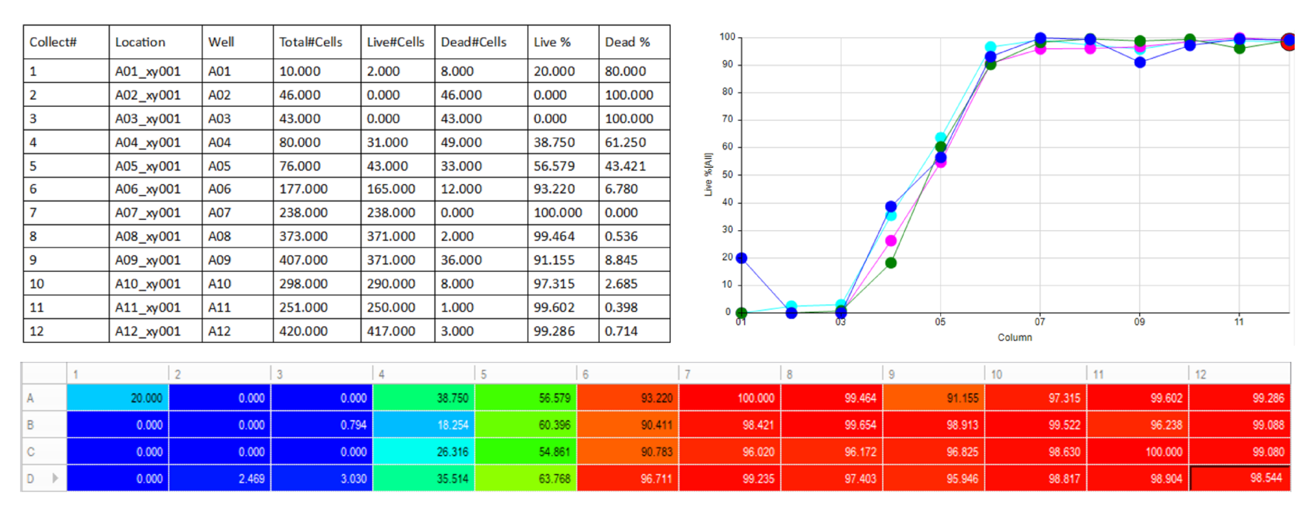
Automatically generate tables, heat maps, charts and even complex bespoke reports.
Measurement parameters supported
Required Modules
Recommended Package
Apps are designed for customers with a registered copy of Media Cybernetics software. Please review the App Center specifications tab to ensure your software is supported. To download the App, please fill out the form below.
Thank you for providing your information.
Please download the App using the link below.
Adding {{itemName}} to cart
Added {{itemName}} to cart