Wound Healing (Scratch Assay)
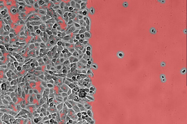
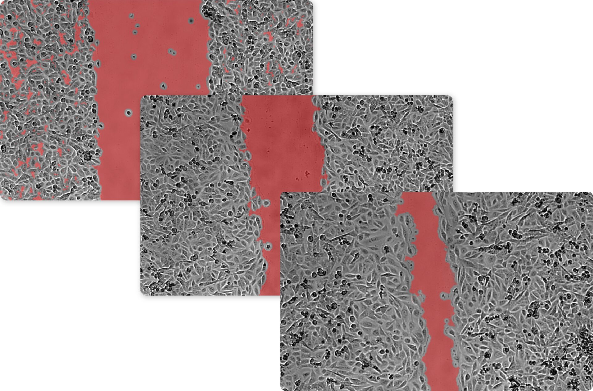
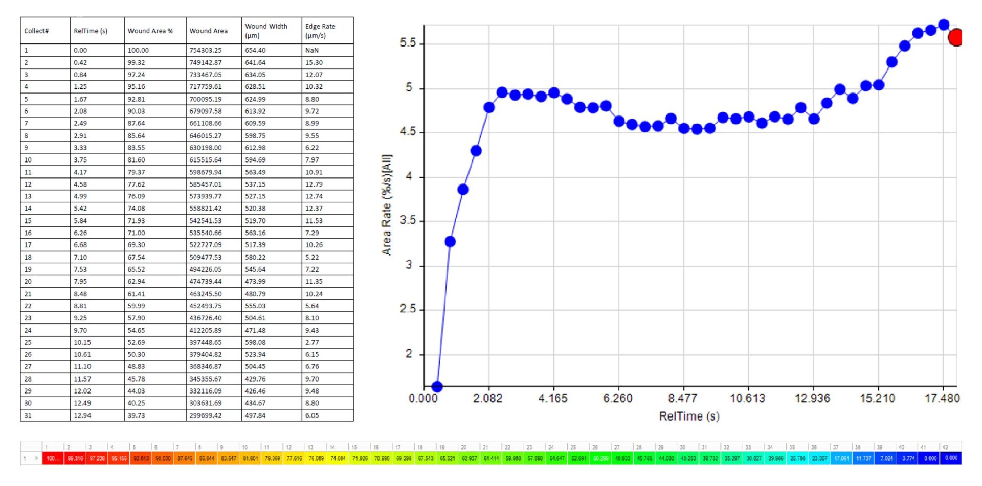
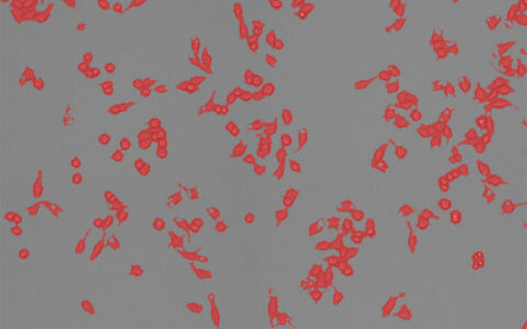
The wound healing assay, indispensable in cancer research, developmental studies, and tissue repair, is a standard in vitro technique for studying collective cell migration in two dimensions. It involves creating a cell-free area in a confluent monolayer through physical exclusion or mechanical, thermal, or chemical damage. This "sheet migration," typical of epithelial and endothelial monolayers, maintains intercellular junctions (Jonkman et al. 2014).
Often called a ‘scratch assay,’ it is performed by scratching a cell monolayer (e.g., with a pipette tip) and capturing time-lapse images. Simple, inexpensive, and widely used, it models cell movement under controlled conditions (Grada et al. 2017). The Image-Pro Wound Healing protocol offers an accurate and reproducible solution for analyzing assay data.
Techniques: Brightfield
How it works
Name Scratch Region
Name the scratch region of your image.
Segmentation
Apply and adjust segmentation to match the intensity of the scratch region.
Time-Lapse
Apply segmentation to all frames of time-lapse.
Quantitative results
Automatically generate tables, heat maps, charts and even complex bespoke reports.
Measurement parameters supported
- • Cell-Free Area Percentage
- • Wound Width (μm)
- • Area Rate (%/s)
- • Edge Rate
- • Time to 50%
- • Custom user-defined measurements
Solution requirements
Required Modules
Base
2D Automated Analysis
Cell Biology Plus Protocol Collection
Wound Healing Protocol
Recommended Package
Literature spotlight
- Liu, Z., Yang, J., Chen, Y., Chen, C., Wang, J., Lee, Y. M., ... & Luo, G. (2022). P311 facilitates the angiogenesis and wound healing function of MSCs by increasing VEGF production. Frontiers in Immunology, 13, 821932.
- Yixiu, Z., Wang, X., Yang, S., Song, X., Chen, C., Sun, N., Zhang, Y., Yao, D., Huang, J., Wang, J. and Zhang, Y., 2020. Kanglexin promotes diabetic wound healing through activating FGFR/ERK1/2 signaling pathway mediated angiogenesis.
- Zhang, P., Lai, X., Zhu, M.H., Long, M., Liu, X.L., Wang, Z.X., Zhang, Y., Guo, R.J., Dong, J., Lu, Q. and Sun, P., 2021. Saikosaponin A, a triterpene saponin, suppresses angiogenesis and tumor growth by blocking VEGFR2-mediated signaling pathway. Frontiers in pharmacology, 12, p.713200.