Cell Proliferation
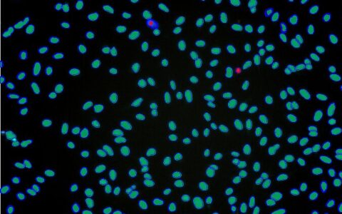
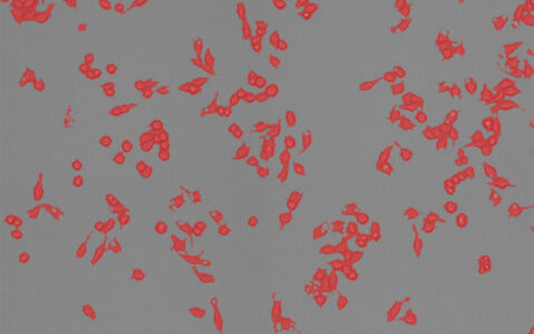
Cell proliferation, the process by which a cell grows and divides to produce two daughter cells, is fundamental to understanding tumorigenesis, where cell cycle disorganization leads to uncontrolled proliferation (Golias et al., 2004).
Proliferation assays are commonly used to evaluate cellular responses to drugs or chemical agents (Adan et al., 2016). These assays often involve the mitotic index or labeling indices using DNA synthesis markers, such as bromodeoxyuridine or tritiated thymidine, or intrinsic markers like Ki67 and proliferative cell nuclear antigen (PCNA) (Goodland, 2017).
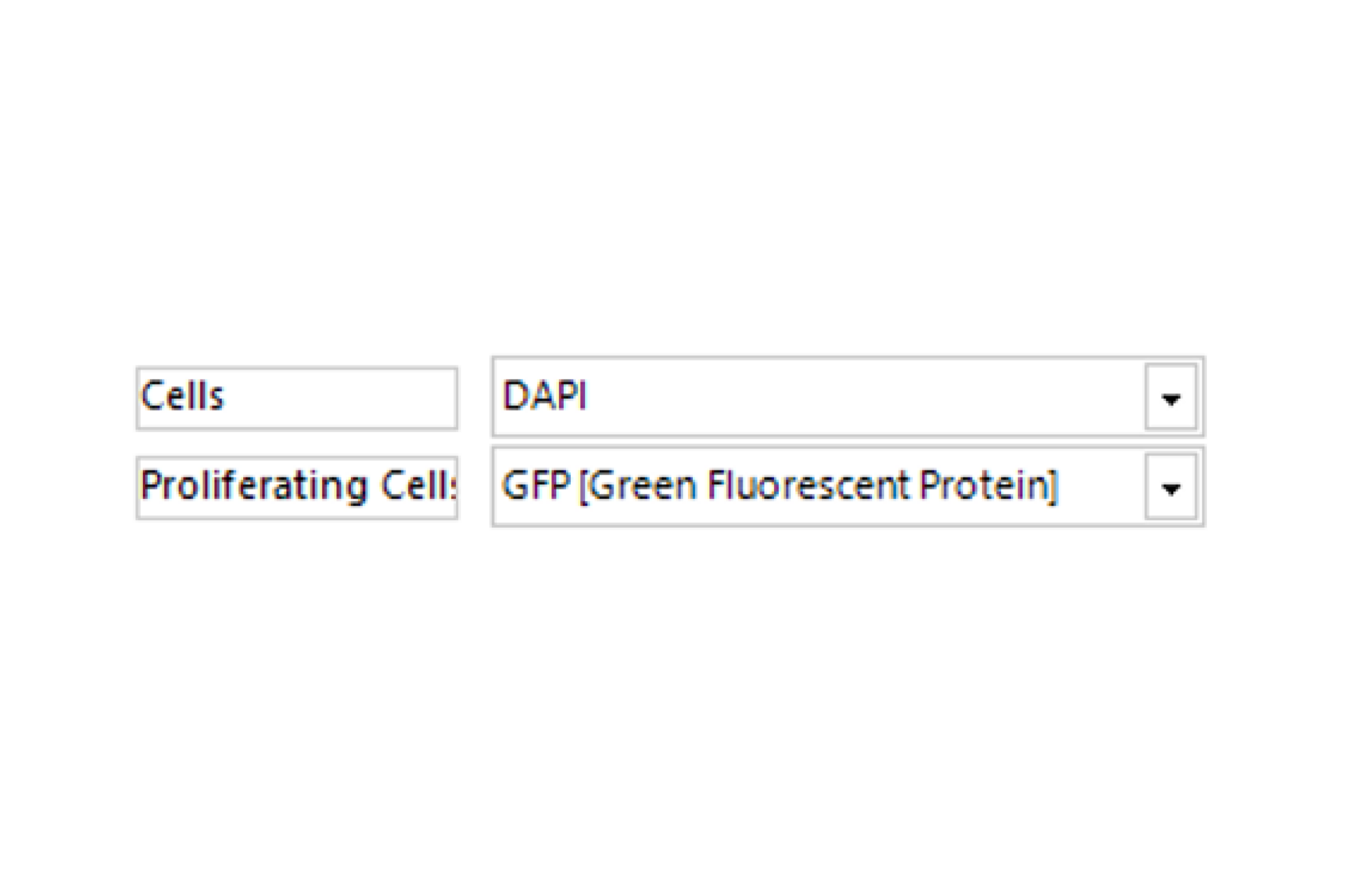
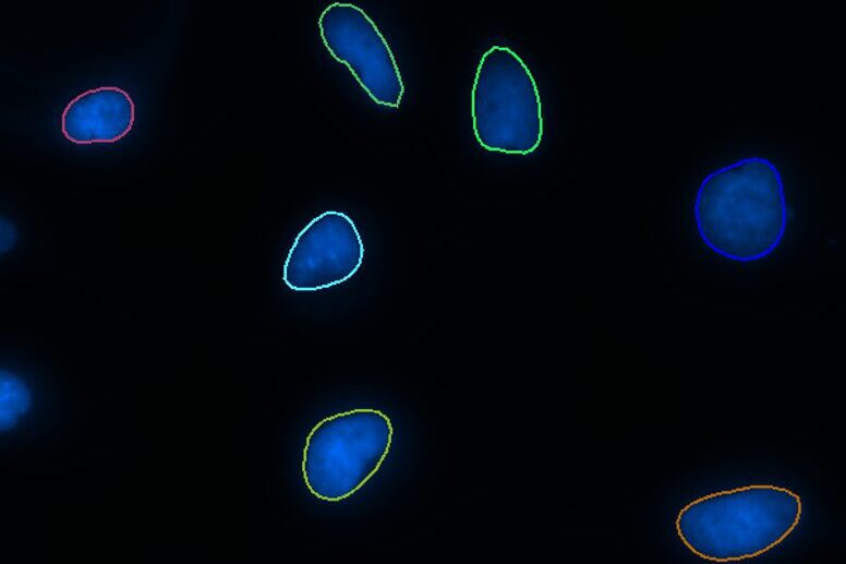
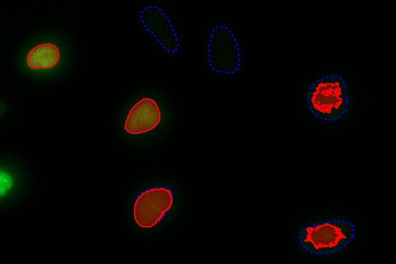
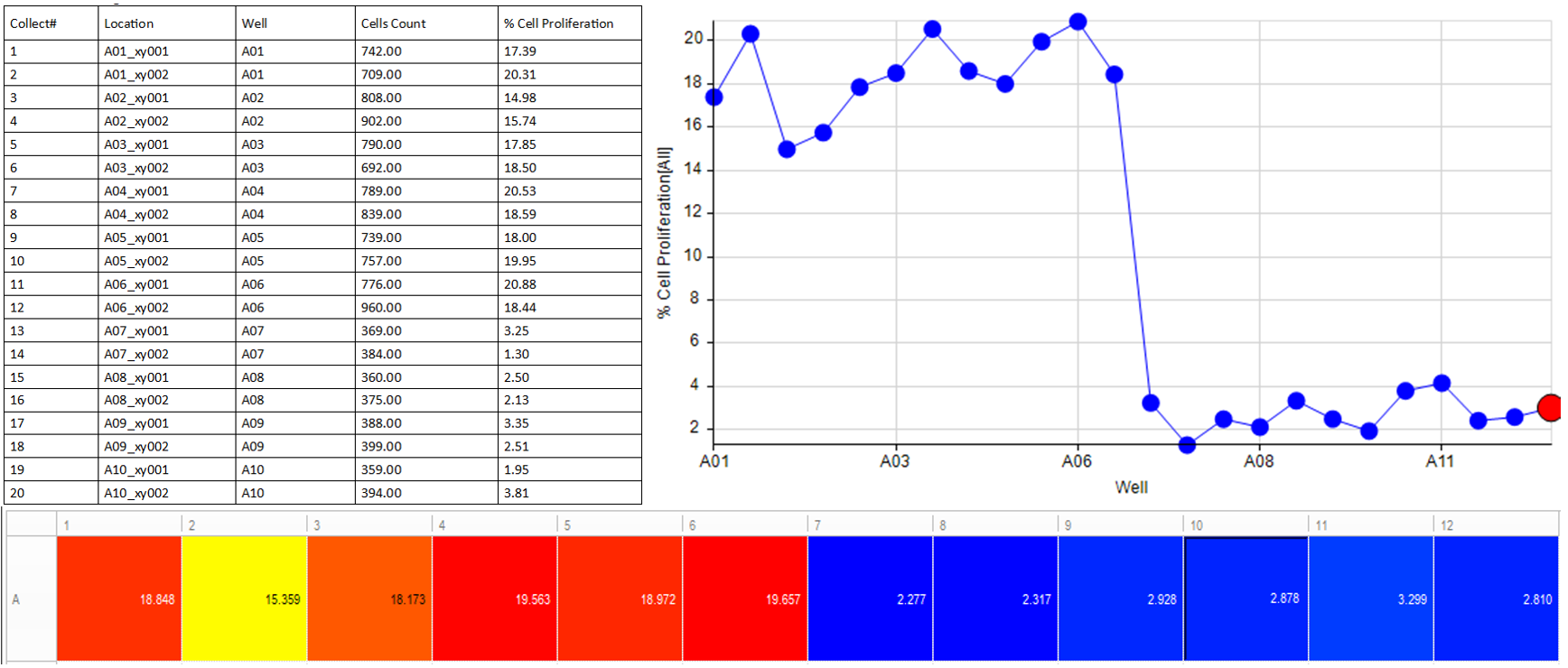
The Image-Pro Cell Proliferation protocol is an ideal tool for analyzing these assays, enabling efficient processing of large datasets, including complex formats like multi-well plates. Its intuitive design ensures accessibility for users with little to no image analysis experience.
Techniques: Fluorescence
How it works
Select Channel
Select the channels that contain labelled cells (or nuclei) and the channel that contains the proliferation marker.
Find Cells or Nuclei
Find cells or nuclei with a pre-trained deep learning model, machine learning or threshold segmentation.
Separate from Background
Separate the proliferation marker from the background with either threshold segmentation or machine learning.
Quantitative results
Automatically generate tables, heat maps, charts and even complex bespoke reports.
Measurement parameters supported
- • Cell Count
- • Proliferating Cell Count
- • Proliferating Cell Percentage
- • Custom user-defined measurements
Solution requirements
Required Modules
Base
2D Automated Analysis
Cell Biology Protocol Collection
Cell Proliferation Protocol
AI Deep Learning
Life Science Models
Fluorescent Cells Model
Recommended Package