Skip links

New App Layout Example

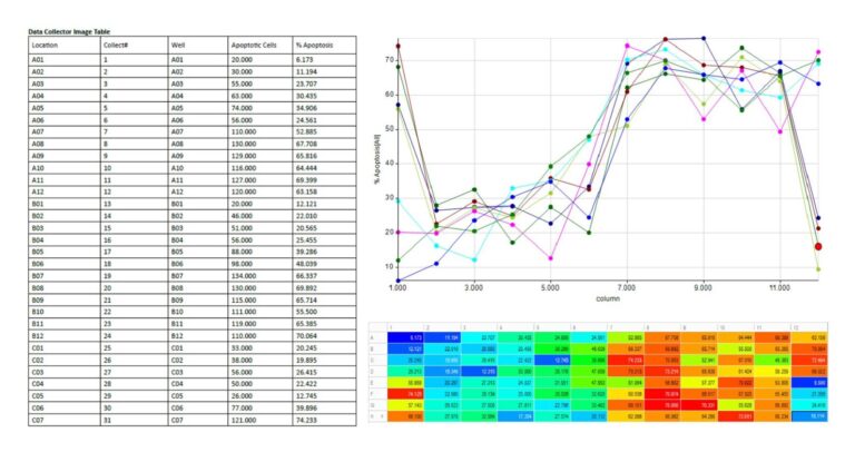
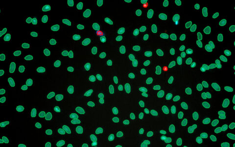
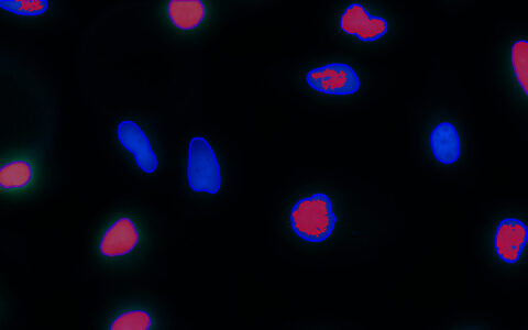
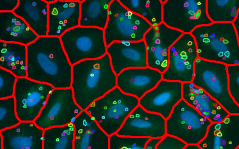
Apoptosis is programmed cell death seen in metazoans and some single-celled eukaryotes like yeast. It plays a vital role in processes such as cell turnover, immune response, development, and hormone-dependent atrophy, and in chemical-induced cell death (Lawen 2003). The failure to initiate apoptosis in mutated cells contributes to cancer, making its analysis crucial in cancer research. Morphological changes during apoptosis include cell separation, membrane structure loss, and blebbing (Wyllie 1997). Caspase proteins are key to apoptosis and are used in assays to detect apoptotic cells. Image-Pro Apoptosis protocol simplifies large-scale analysis of these cells, even for users with minimal image analysis experience.

Techniques: Fluorescence

How it works

Raw Image

Select the channel that contains labeled nuclei and the channel that contains labeled apoptosis marker.

Segment Nuclei

Find nuclei with a Pre-Trained Deep Learning Model, Machine Learning, or Threshold Segmentation.

Segment Marker

Find the apoptosis marker with a Pre-Trained Deep Learning Model, Machine Learning, or Threshold Segmentation.

Quantitative results

Automatically generate tables, heat maps, charts and even complex bespoke reports.

Measurement parameters supported

Solution requirements

Required Modules

Base

2D Automated Analysis

Cell Biology Protocol Collection

Cell Apoptosis Protocol

AI Deep Learning

Life Science Models

Fluorescent Cells Model

Literature spotlight

RELATED APPS

Request a demo to see how it works